Plant Cell Diagram Under Light Microscope / Cell Structure Teaching Resources The Science Teacher / Cells of plant or animal tissue.. It also has a very high resolving power. The diagram below is a plant cell as may be seen using a light microscope. Light photomicrograph of helianthus stem cross section seen through microscope. To use a light microscope to examine animal or plant cells. Under a light microscope, the cell membrane, nucleus and cytoplasm of a cheek cell (animal cell) can be observed.
Plant cells are the basic unit and building blocks of life in organisms of the kingdom plantae. Transport proteins modified by the golgi body outside of the cell. We say cells are microscopic because they can only be seen under a microscope. See how a generalized structure of an animal cell and plant cell look with labeled diagrams. The diagram below is a plant cell as may be seen using a light microscope.
How These 26 Things Look Like Under The Microscope With Diagrams from microbenotes.com Plant cells are the basic unit and building blocks of life in organisms of the kingdom plantae. Purple colored, large epidermal cells of an onion oyster plant cells. Microscopy and the interpretation of cell structures. Describe and compare the structure of a plant cell with an animal cell, as seen under a light microscope, limited to cell wall, nucleus, cytoplasm, chloroplasts, vacuoles and location of the cell membrane. The diagram below is a plant cell as may be seen using a light microscope. Resolving power is the ability to distinguish between separate things which are close to each other. To make observations and draw scale diagrams of cells. A cell is a very tiny structure which exists in living bodies.
Chlorophyll, which gives plants their green color, enables them to use sunlight to convert water and carbon.
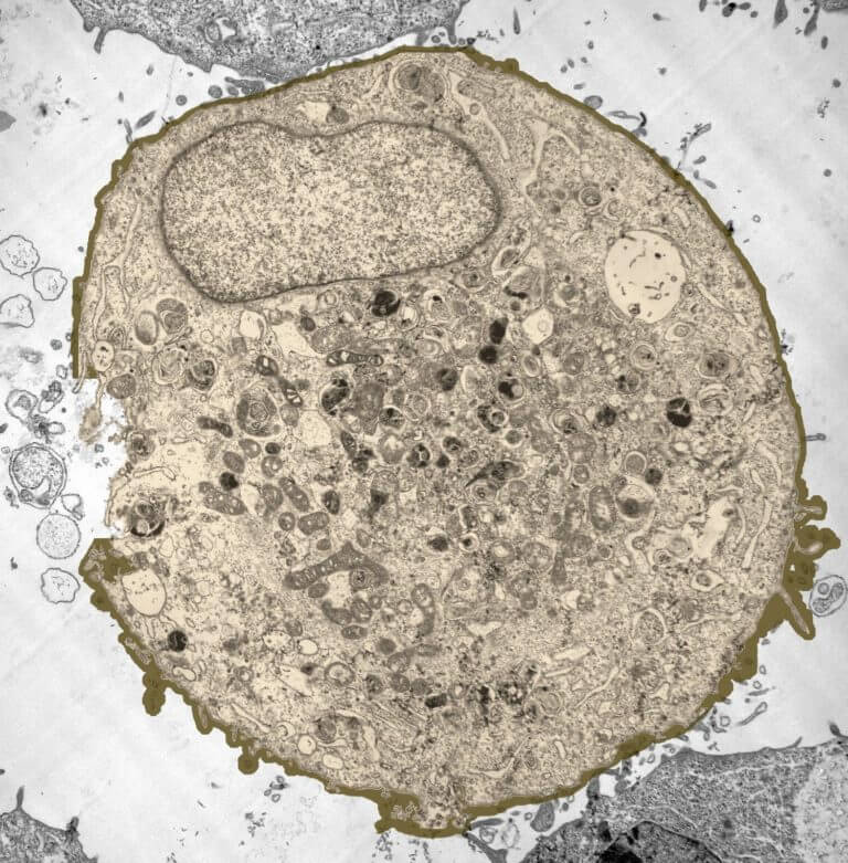
Animal cells also have a many of the differences between plant and animal cells are visible under a microscope, and it's relatively straightforward to distinguish between the two. Turn the coarse focus so that the stage is as close to the objective lens as possible. See how a generalized structure of an animal cell and plant cell look with labeled diagrams. Cells of plant or animal tissue. A few cell organelles can be seen when a plant cell is viewed under a light microscope. Here's a photo of a plant cell under an electron microscope. Plant cell is an eukaryotic cell primarily involved in photosynthesis and having its genomic content some of these differences can be clearly understood when the cells are examined under an electron microscope. General animal cell as seen under the light microscope. Tulip stem cells at the microscope. Make sure you can label ribosomes and mitochondria on a cell diagram. Juicy green plant cells under the microscope. It also has a very high resolving power. A scale bar has been marked on the drawing, allowing the size of a cell to be estimated.
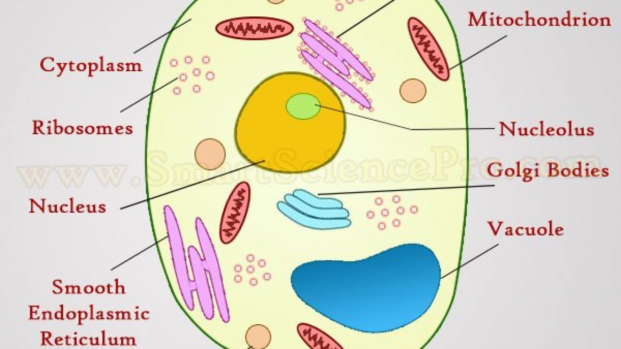
Study the two diagrams of plant and animal cells below. Plant cells have cell walls, one large vacuole per cell, and chloroplasts, while animal cells will have a cell membrane only. Structure of animal cell and plant cell under microscope + diagrams. Diagram 1 shows the structure of a plant cell as seen under light microscope. The plant cells under microscope plant cells under microscope.
Cells Mr Wong S Class Website from www.wjfclass.net A few cell organelles can be seen when a plant cell is viewed under a light microscope. The high resolving power makes the electron microscope a very important research tool in microbiology. Plant cell is an eukaryotic cell primarily involved in photosynthesis and having its genomic content some of these differences can be clearly understood when the cells are examined under an electron microscope. Here's a photo of a plant cell under an electron microscope. Just place your prepared slide of plant between light and slide stand and focus on 40x or 100x you can easily see plant cells under microscope. Cell is a tiny structure and functional unit of a living organism containing various parts known as organelles. Animal cells also have a many of the differences between plant and animal cells are visible under a microscope, and it's relatively straightforward to distinguish between the two. Tulip stem cells at the microscope.
Use them in commercial designs under lifetime, perpetual & worldwide rights.
Resolving power is the ability to distinguish between separate things which are close to each other. Under a light microscope, the cell membrane, nucleus and cytoplasm of a cheek cell (animal cell) can be observed. You should not look through the microscope to do this. A cell is a very tiny structure which exists in living bodies. Plant cells are eukaryotic cells present in green plants, photosynthetic eukaryotes of the kingdom plantae. See how a generalized structure of an animal cell and plant cell look with labeled diagrams. Under ordinary light microscope only few cell organelles like mitochondria, golgi complex however, under electron microscope, several other cytoplasmic organelles such as endoplasmic plastids are present only in plant cells (not in animal cells). A few cell organelles can be seen when a plant cell is viewed under a light microscope. The inner layer is continuous and forms flattened membrane sacs called thylakoids. The diagram is very clear, and labeled; Cells of plant or animal tissue. Turn the coarse focus so that the stage is as close to the objective lens as possible. They carry out aerobic respiration producing atp.
Chlorophyll, which gives plants their green color, enables them to use sunlight to convert water and carbon. It also has a very high resolving power. Turn the coarse focus so that the stage is as close to the objective lens as possible. A micrograph is a photo or digital image taken through a microscope to show a magnified image of a specimen. Plant cells have cell walls, one large vacuole per cell, and chloroplasts, while animal cells will have a cell membrane only.
Structure Of Animal Cell And Plant Cell Under Microscope Diagrams from www.smartsciencepro.com Light photomicrograph of helianthus stem cross section seen through microscope. Plant cells have cell walls, one large vacuole per cell, and chloroplasts, while animal cells will have a cell membrane only. Onion epidermis under light microscope. A few cell organelles can be seen when a plant cell is viewed under a light microscope. Light microscope slide with microsection of an evergreen conifer in. Juicy green plant cells under the microscope. It also has a very high resolving power. General animal cell as seen under the light microscope.
The diagram is very clear, and labeled;
Under a light microscope, the cell membrane, nucleus and cytoplasm of a cheek cell (animal cell) can be observed. The diagram below is a plant cell as may be seen using a light microscope. To make observations and draw scale diagrams of cells. Just place your prepared slide of plant between light and slide stand and focus on 40x or 100x you can easily see plant cells under microscope. Microscopy and the interpretation of cell structures. Vector low poly solar power plant and city. Tulip stem cells at the microscope. Each part, known as an organelle, works together to keep the cell functional. Transport proteins modified by the golgi body outside of the cell. Light photomicrograph of helianthus stem cross section seen through microscope. These include the cell wall, cell membrane, nucleus, chloroplasts. Purple colored, large epidermal cells of an onion oyster plant cells. But at the same time it is interpretive.


0 Comments